A Patient’s Question During Checkups: How Often Should You Get Dental X-Rays?
When patients sit down for a routine checkup at SmileNote, one of the questions that often comes up is surprisingly simple: how often should you get dental X-Rays?
This topic frequently appears in discussions about preventive dental care. Many people assume that X-rays are automatically taken at every dental visit, but the reality is more nuanced. Understanding how often should you get dental X-Rays usually begins with a conversation between a dentist and a patient about oral health history, current symptoms, and the purpose of dental imaging.
What Dental X-Rays Actually Show
During a dental examination, dentists visually inspect the teeth, gums, and soft tissues. However, certain problems develop in areas that cannot be seen directly.
Diagnostic Capabilities
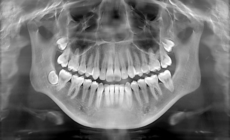
Dental X-rays help reveal:
- Cavities between teeth
- Infections near tooth roots
- Bone loss around teeth
- Impacted teeth
- Hidden structural damage
Without radiographs, these conditions could progress quietly until discomfort or visible damage appears.
Individualized Imaging Schedules
Patients sometimes expect a standard timeline, such as “once per year.” In practice, dentists rarely apply identical imaging schedules to every patient.
Some individuals have very stable oral health and may not require frequent radiographs. Others maintain excellent oral hygiene and rarely develop cavities. In these cases, dentists may extend the interval between radiographs because the likelihood of hidden dental disease is relatively low.
Conversely, some patients experience recurrent cavities or gum disease and may benefit from more regular imaging. Therefore, when discussing how often should you get dental X-Rays, dentists typically evaluate each person’s dental history.
Situations Requiring More Frequent Imaging
Dentists sometimes recommend additional X-rays when specific conditions are present. Examples include:
- New dental pain or tooth sensitivity
- Signs of swelling or infection
- Treatment planning for procedures such as implants or orthodontics
- Monitoring healing after certain treatments
Although imaging is not always required, it can provide valuable diagnostic information when symptoms appear.
Addressing Concerns About Radiation and Communication
Safety Protocols
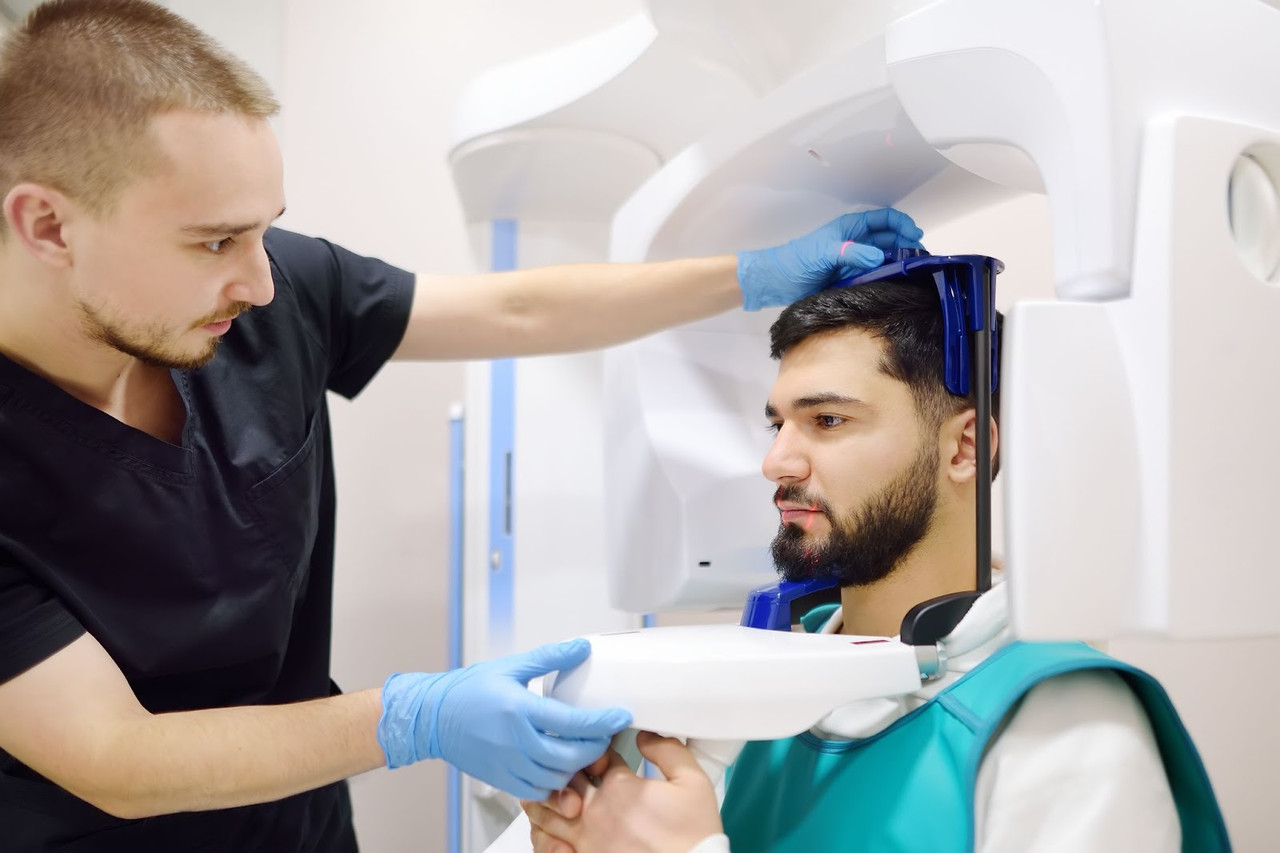
Many patients naturally ask whether dental X-rays are safe. Modern dental imaging systems use very small radiation doses. Protective measures such as lead aprons and digital sensors further reduce exposure. According to information summarized by organizations such as the American Dental Association and the NHS, dental radiographs are considered safe when used appropriately as part of diagnostic care. Dentists always aim to take images only when they provide meaningful clinical benefit.
One of the most helpful things patients can do is simply ask questions during their appointment. Understanding the reason behind imaging recommendations helps patients feel more comfortable with their treatment plan. When dentists explain imaging frequency, they are not just discussing technology—they are describing how imaging fits into a larger strategy of preventive oral health care.
Conclusion
The question how often should you get dental X-Rays does not have a single universal answer. Instead, imaging frequency depends on factors such as oral health history, current symptoms, and the dentist’s clinical evaluation.
For many patients, dental X-rays are an important diagnostic tool that allows problems to be detected early, often before symptoms develop. Discussing imaging decisions with a trusted dental professional ensures that care is tailored to individual needs while maintaining safety and effectiveness. Ultimately, the diagnosis based on your unique biological and mechanical factors will determine exactly how often should you get dental X-Rays.