Microstructural Failure Patterns: Can You Put a Crown on a Broken Tooth?
To refine the clinical answer to restorative predictability, this SmileNote analysis examines microstructural behavior at the enamel-dentin interface. Fractures rarely propagate randomly. Instead, they follow lines of stress concentration influenced by enamel rod orientation and dentinal tubule direction.
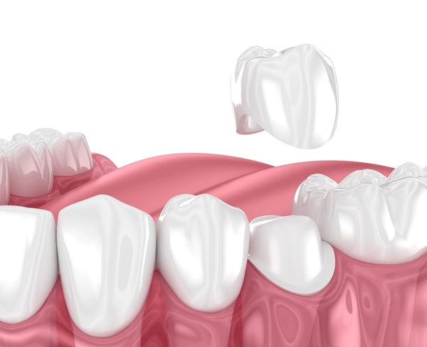
Microstructural Failure Patterns in Fractured Teeth
Enamel is highly mineralized and brittle. Dentin is resilient and capable of limited flexure. When excessive occlusal force or trauma occurs, stress often concentrates at:
- Marginal ridges
- Developmental grooves
- Sites of previous restorations
- Areas of internal caries undermining enamel
Fracture Propagation
If fracture propagation terminates within dentin and does not cross the cemento-enamel junction, restorative predictability increases. However, once cracks extend apically along dentinal tubules toward the root surface, structural prognosis declines. Crown placement does not eliminate crack propagation risk. It redistributes force but cannot biologically “heal” internal microfractures. This distinction is critical in structural assessment.
.webp)
Adhesive Interfaces and the Limitations of Bonding
Modern adhesive systems have improved dentin bonding reliability. However, adhesive strength must be evaluated relative to functional load.
Macro-Retention vs. Adhesion
Bonded restorations depend on hybrid layer formation, resin tag penetration, and micromechanical interlocking. While adhesive dentistry allows conservative management in some fractured teeth, full-coverage crowns rely less on bonding and more on macro-retention (axial wall height and taper). When residual tooth structure is minimal, reliance solely on adhesive retention becomes biomechanically insufficient.
Clinical data suggest that adhesive reinforcement can support fractured cusps in select cases, but when more than 50% of coronal structure is compromised, indirect full coverage often provides more predictable load distribution. Thus, the feasibility of crown therapy remains structurally dependent rather than adhesive-dependent.
Periodontal Interface Stability
Any discussion of restorative success must integrate periodontal dynamics. A stable crown requires healthy gingival attachment, controlled sulcus depth, absence of active periodontal inflammation, and adequate keratinized tissue.
If fracture margins extend into the supracrestal tissue attachment zone, surgical crown lengthening may be indicated. This procedure re-establishes proper biologic spacing between the restorative margin and alveolar bone crest. However, crown lengthening reduces the clinical crown-to-root ratio. Therefore, the intervention must balance restorative accessibility with long-term periodontal stability. In compromised periodontal environments, extraction and prosthetic replacement may provide more predictable outcomes.
Endodontically Treated Teeth and Fracture Susceptibility
Teeth requiring root canal therapy prior to crown placement demonstrate altered structural behavior. Contrary to common belief, endodontic treatment itself does not inherently weaken dentin. Structural compromise arises primarily from loss of coronal tissue during access preparation and caries removal.
- Reduced proprioception
- Increased cusp flexure when inadequately restored
- Elevated fracture risk without full coverage
Multiple longitudinal analyses demonstrate improved survival rates when posterior endodontically treated teeth receive crowns rather than large direct restorations. Therefore, in many post-endodontic fracture scenarios, crown placement is not merely optional—it is protective.
Margin Design and Occlusal Modification
Margin configuration influences stress concentration and periodontal health (e.g., chamfer, shoulder, feather edge). For fractured teeth, margin design must preserve maximal remaining dentin while providing adequate ceramic thickness. Excessive reduction may further weaken structural integrity, while insufficient reduction predisposes to ceramic fracture. Biomechanical modeling indicates that evenly distributed circumferential margins reduce localized tensile stress.
Furthermore, restoring a fractured tooth without evaluating occlusion may result in recurrence. Functional parameters such as maximum intercuspation contacts, lateral excursive guidance, and parafunctional wear patterns must be assessed. Selective occlusal adjustment or fabrication of a protective occlusal guard may be necessary to reduce functional overload.
Synthesis of Clinical Determinants and Conclusion
Integrating biological, mechanical, and functional variables, crown placement is appropriate when the fracture does not extend vertically through the root, adequate ferrule is attainable, periodontal support remains stable, endodontic pathology is controlled, and occlusal forces are managed.
Contraindications for Crown Therapy
Conversely, crown placement is contraindicated when the structural foundation is non-restorable, root integrity is compromised, bone support is insufficient, or repeated fracture risk is high. Thus, the feasibility threshold is measurable and clinically definable.
From a structural standpoint, the restorative solution is resolved through biomechanical assessment rather than optimism. Crowns are protective restorations designed to redistribute force and preserve remaining structure. They are not curative solutions for foundational instability. A broken tooth can often be restored successfully with a crown when anatomical parameters meet established prosthodontic criteria. When those parameters are absent, alternative approaches—including surgical modification or extraction—may provide greater long-term stability.
Clinical judgment must integrate fracture classification, periodontal biology, pulpal vitality, occlusal load, and material science. Only when these variables align does crown therapy offer predictable longevity and confidently answer whether can you put a crown on a broken tooth.